Hip Joint
Pathology of the Hip joint may refer symptoms into the groin. Click on the following links to learn more about each pathology.
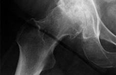
Avascular Necrosis of the Femoral Head
Avascular necrosis, most commonly seen the femoral head, results when the vascular supply of the bone is interrupted leading to bone death. Many times, the necrosis is idiopathic, asymptomatic, and the cause of it is still not clearly understood by the medical community. However, early detection is important in order to reduce the collapse of the articular surface which leads to degeneration of the hip. In very severe cases, a joint replacement may need to be done in order to repair the dead area. This condition is serious and should be referred out to a physician.
Slipped Capital Femoral Epiphysis
Slipped Capital Femoral Epiphysis (SCFE) is caused by a “posterior and inferior slippage of the proximal femoral epiphysis on the metaphysis (femoral neck), which occurs through the epiphyseal plate”. This condition usually occurs in children between the ages of 8 and 15 years of age, usually occurs in boys more than girls, in children that are obese, and is more prevalent in African Americans and Pacific Islanders. The prognosis of SCFE is largely related to how quickly the condition is diagnosed and treated. If the it is not caught early, which happens often due to vague symptoms, it can lead to early-onset degenerative hip arthritis that may eventually lead to a hip reconstruction. Treatment usually includes crutches or a wheelchair and possible surgery to prevent further slippage. If the therapist suspects this condition, the child should be referred in order to get a radiography to confirm the diagnosis.
Osteoarthritis of the Hip
Osteoarthritis (OA) of the hip is one of the most common joint disorders that affects the joint and causes capsular changes within. OA usually occurs in middle age and elderly people and causes cartilage loss, subchondral bone and joint degeneration, decreased joint space, flattening of the formal head, and a shortening of the joint capsule. Over time, osteophyte formation may occur from the excessive tensile forces on the capsule or from the increased pressure on the articular cartilage. This disease causes loss of range of motion (especially internal rotation and flexion), stiffness, and muscle weakness especially of the abductors. These changes associated with the disease are usually slow to progress but in some cases can happen very rapidly but the disease process can be slowed through proper exercise and management. Treatment of OA includes patient education on ways to decrease pain, improve their function, reducing stiffness, and reducing the need for medications, functional gait and balance training, the use of an assistive device, manual therapy, strengthening exercises, and flexibility exercises. However, some individuals may require a total hip arthroplasty if they do not respond to treatment.
Hip Dislocation/Subluxation
Hip dislocations and subluxations are common in athletes and older adults, especially ones who have just undergone a total hip arthroplasty (THA), and can happen as a result of trauma. A hip dislocation occurs when the femoral head comes out of the acetabulum and a subluxation occurs when the femoral head does not completely translate out of the acetabulum. While it is very uncommon for the hip to completely dislocate, subluxations are more frequent but are inadequately recognized. Most of the hip dislocations are in the posterior direction, while anterior dislocations account for only 8% to 15% of the total dislocations. Dislocations and subluxations can occur by falling onto a flexed knee with the hip adducted or if the individual stops quickly and pivots on their weight bearing extremity. Individuals who have undergone a THA with a posterolateral approach are also more inclined to suffer a hip dislocation if the patient does not abide by the hip precautions. Individuals with a posterior dislocated hip usually present with a flexed, adducted, and internally rotated leg; whereas individuals with an anterior dislocation present with a leg that is externally rotated, abducted, and either flexed or extended. Treatment includes reducing the dislocated hip and then if there are no other complications, such as a fracture, the individual will be put on crutches until they are able to walk without pain. Physical therapy is also important to regain strength and normal function in the involved joint. This is a serious injury and the patient should be referred to a doctor in order to decrease complications or more injury to the joint and surrounding structures. Dislocations and subluxations many times result in fractures to the posterior acetabulum or the femoral so the individual should be referred for imaging.
Red Flags
References
1. Karapinar L, Gurkan A, Kacar S, Polat O. Post-transplant femoral head avascular necrosis: a selective investigation with MRI. Ann Transplant 2007;12:27-31.
2. Goodman C, Snyder T. Differential Diagnosis for Physical Therapists: Screening for Referral. 4th ed. St. Louis: Suanders Elsevier; 2007.
3. Peck D. Slipped capital femoral epiphysis: diagnosis and management. Am Fam Physician 2010;82:258-62.
4. Cibulka MT, White DM, Woehrle J, et al. Hip pain and mobility deficits--hip osteoarthritis: clinical practice guidelines linked to the international classification of functioning, disability, and health from the orthopaedic section of the American Physical Therapy Association. J Orthop Sports Phys Ther 2009;39:A1-25.
